
![]()
Anthropology report phase III of the excavations at the Islamic necropolis in Tauste.

12 exhumed skeletons: 9 adults and 3 children
The El Patiaz Cultural Association from Tauste (Zaragoza) has conducted a new trial excavation on the Obispo Conget Avenue as a part of the Anthropology report of the Muslim necropolis found earlier in the town. These works are meant to continue the research started after the excavations carried out two years ago.
This time, 12 skeletons were exhumed, most of them incomplete. They correspond to 9 adults and 3 children. Similarly to the remains exhumed in the previous excavations, the burial types as well as the orientation of the bodies, match the patterns of the Islamic necropoleis: the bodies face the Mecca, they lie on their side (lateral decubitus) and bear no funerary objects.
Nine out of twelve skeletons uncovered correspond to three women and six men, all adults and generally middle-aged, only one of them would be younger than 40. All nine share a noticeable oral pathology: abscesses, fistulas, dental caries, plaque and tartar build-up. Signs of osteoarthritis, especially in the vertebral bodies, can be observed in most cases. There are clear signs of physical activity, such as arm bending, squatting, and having long walks.
As for the skeletons of children, two of them were as young as 2 years old and the third would be slightly older, between 2 and 4.
During the excavations, it was found that the tombs had two levels. One of the skeletons comes from a deeper and therefore older layer. The remains belong to a man aged between 25 and 35. This skeleton is one of the oldest among those found and as a consequence is not well preserved. It was seriously deteriorated by land loads and pressure.
Trepanning.
One of the most relevant features of the exhumed remains is the sign of trepanning found on one of the male skeletons aged around 45. It must have been the hole-drilling trepanning procedure. The mark of the wound can be appreciated on the surface of the skull.
The regeneration of the skull bone indicates that the patient survived the surgery, although there are also signs of the subsequent infection he might have suffered. The trepanning technique has been practiced since ancient times, and its presence indicates an intervention with either medical or ritual purposes.
In light of the results of this analysis that has been conducted on the remains extracted during the trial excavations in question, we can conclude that all the skeletons found, belong to the Caucasian race and share certain pathologies. The periodontal disease, in particular, was very noticeable in most skeletons of adult individuals who had signs of significant tooth wear caused, on the one hand, by intense mastication and, on the other, by lack of oral and dental hygiene back in the day.
ANTHROPOLOGICAL ANALYSIS
Anthropology study made it possible to identify the sex, age and height of most preserved skeletons. This is the summarized description of the exhumed remains and their features observed as a result of the above-mentioned study.
3 – Skeleton – sex: male;
Height: between 1.60 and 1.65 meters; age: approximately 45 years old. The most relevant feature: signs of trepanning procedure on the left parietal lobe, apparently executed by drilling a circle of holes; an incision groove made by the tool. Edges with beveling indicate regrowth of the bone on the perimeter, which means it healed while the patient was alive. The porosities around the trepanned area indicate some kind of subsequent infection that might have been caused by the wound and correspond to the osteomyelitis lesion. The abscesses found on the femoral and tibia heads are compatible with osteomyelitis.
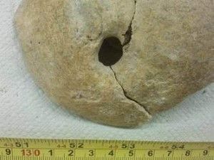

Skull trepanning by hole drilling, followed by patient’s survival. Trepanning and possible osteomyelitis lesion.
1 – Skeleton – sex: female;
Height: 1.60 meters; age: between 45 and 60. The deterioration of the bones is compatible with old age. Some signs of osteoarthritis can be appreciated in the spinal vertebrae. There are also some indications of phalanx and ulnar enthesopathy that might be attributed to the intense activity of the upper limbs. One can also appreciate the presence of periodontal disease, as well as caries, plaque build-up and enamel hypoplasia.


2 – Skeleton- sex: male;
Only the lower part of the skeleton is preserved, height: ranging between 1.60 and 1.62 meters. The age of these remains cannot be determined but the deterioration of the bones is compatible with old age. Some signs of osteoarthritis can be appreciated in the femoral neck, as well as a facet of the femoral condyle that is associated with repetitive squatting.

4 – Skeleton – sex: male;
Estimated height: between 1.58 and 1.62 meters; age: between 40 and 45. It has signs of periodontal disease, elastic deformation of the alveolar bone and tooth wear. Condition of the bones of the upper limbs reveals that the individual was exposed to significant physical activity. There are also some signs of osteoarthritis in the vertebrae and spurs on both patella bones that are associated with walking and squatting.


Osteophytes in the heel and patella bones. Upper dental arch showing the elastic deformation of the alveolar bone as a result of the periodontal disease.
5 – Skeleton – sex: male;
Approximate height: ranging between 1.60 and 1.65 meters; age: between 40 and 50. Similar to the previous skeleton, this one has some noticeable indications of dental disease and vertebral pathology that shows vertebral crush and herniation.


7 – Skeleton – sex: female;
Height: 1.58 meters; age: between 33 and 45. It also shows signs of oral pathologies, such as caries and tartar build-up, as well as osteoarthritis in the metacarpal bones and lumbar vertebrae.

9 – Skeleton- sex: male;
With extremely robust bones that belong to a male individual whose approximate height ranged between 1.72 and 1.76 meters; age: ranging between 33 and 45. Paleopathological analysis suggests intense activity associated with elbow flexion. The deterioration of the bones is compatible with old age. The remains have signs of osteoarthritis in the vertebrae and both femoral heads.

10 – Skeleton – sex: male;
Only an upper limb of the body is preserved, height: ranging between 1.62 and 1.65 meters; age: between 25 and 35 years old. The body has oral pathologies: fistulas and caries, vertebral wear and spinous process deviation that might point at possible scoliosis.

11 – Skeleton – sex: female;
Height: impossible to estimate; age: ranging between 33 and 45. Similar to other skeletons that belong to old aged individuals, one can observe in this one lumbar crush fracture and phalanx enthesopathy caused by intense physical activity of the limbs. The most noticeable pathology is oral, including caries and tooth wear leading to erosion and loss of the crown. One can also appreciate the presence of an ectopic (supernumerary) tooth.

6 – Skeleton of a child – sex: unidentified; height: approximately 98 centimeters; age: ranging between 2 and 4 years.
8 – Skeleton of a child – sex: unidentified; height: unidentified. Dental analysis shows that it might be approximately 2 years old.
12 – Skeleton of a child – sex: unidentified; height: unidentified. The youngest body uncovered in this excavation: dental analysis shows that its age might range between 18 months and 2 years.
This analysis complements the one that was done in 2011 after the second trial excavation conducted in the same area of Tauste. The goal of the El Patiaz Cultural Association is to continue its work on promoting further research and study of the Muslim necropolis found in Tauste that clearly covers a large area.


 Licencia Creative Commons Atribución-NoComercial-CompartirIgual 3.0 Unported.
Acknowledgement
The translation of this page is included in the Zaragoza Provincial Council Grants for the Dissemination and Revitalisation of Cultural Heritage in the year 2022.
La traducción de está página esta incluida dentro de las Ayudas de la Diputación Provincial de Zaragoza para la Difusión y Dinamización del Patrimonio Cultural en el año 2022.